

MRI scans
Moving Forward to Advanced Imaging
Your neurologist will refer you for an MRI scan. As MS is difficult to diagnose (especially which type), you may have to have a couple… This step is a standard part of narrowing down why you’re experiencing symptoms like foot drop, balance troubles, or sensory changes.
Magnetic Resonance Imaging (MRI) is one of the most powerful tools available in modern medicine. It uses strong magnetic fields and radio waves to create remarkably detailed pictures of the inside of your body without using radiation. For neurological concerns, it lets doctors look directly at your brain and spinal cord to see how signals are travelling.
Why Diagnosing MS Isn’t Straightforward
Multiple sclerosis doesn’t reveal itself through a single simple blood test. Because its symptoms often overlap with dozens of other nerve or spinal conditions, doctors must follow strict diagnostic guidelines to confirm it.
- The Rule of Dissemination… Doctors need proof that nerve damage has occurred in more than one area of the central nervous system and at different points in time.
- Ruling Out Alternatives… Neurologists must systematically check for and eliminate other explanations, such as vitamin deficiencies, Lyme disease, or simple structural nerve compression.
- Pinpointing the Specific Type… Distinguishing between different forms—such as relapsing-remitting or progressive types—requires observing how the condition behaves over months or even years.
“Diagnosing neurological conditions is like solving a complex puzzle… every scan adds a vital piece to the overall picture,” experts observe.
Why You Might Need More Than One MRI
It’s common for people to wonder why a single scan isn’t always enough. In fact, over 60% of people undergoing investigation for potential MS require repeated imaging sessions over time.
Your initial scan provides a baseline picture. If it shows areas of inflammation or scarring (known as lesions), a follow-up scan a few months later helps doctors see if new lesions have formed, even if you haven’t felt new physical symptoms. Repeating the scan using a contrast dye (called gadolinium) can also help specialists distinguish between old, inactive lesions and active areas of current inflammation.
What to Expect During the Scan
Knowing what happens during an MRI scan can help ease any anxiety you might feel before your appointment…
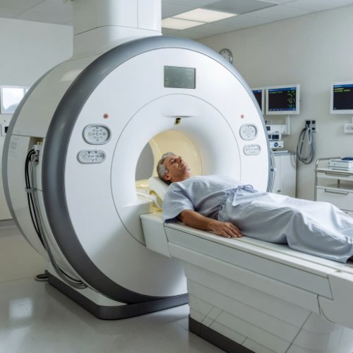
- You’ll lie flat on a motorised bed that slides gently into a large, tunnel-like scanner.
- The machine makes loud tapping and thumping noises as it builds the images, so you’ll be given earplugs or headphones to wear.
- You’ll need to remain as still as possible while the images are taken to ensure the pictures stay sharp and clear.
- The radiographer operates the scanner from an adjacent room but doesn’t leave you isolated—they can see, hear, and talk to you through an intercom throughout the entire procedure.
What Happens After Your Imaging
Once your scans are complete, a radiologist reads the images and sends a comprehensive report back to your neurologist. Your specialist will then schedule a follow-up consultation to walk you through the results.
Whether the scans show evidence of MS, point toward a trapped nerve, or show clear nerve pathways, having concrete visual data helps your care team shape a treatment and management plan tailored specifically to you. Navigating medical testing can feel uncertain, but each diagnostic step brings you closer to clarity, support, and the right strategies to keep moving safely across the world.